Friday, April 29, 2016

Nandini... What is your senior project?

What a great question! I'd like you all to reach way back in time... not too far... just to March. Remember my first post? It kind of explained what I was doing, but "kind of" doesn't cut it. Hopefully these next few paragraphs will give you all a fuller, better, and deeper understanding.

In my last post, I mentioned that the cellular response to the different radiation methods have yet to be investigated.

The University of Arizona Center for Applied Nanobioscience and Medicine has come of with a preliminary study to evaluate these responses.

The question: Is there a difference in cellular response between high single (1x10Gy) and fractionated dose (5x2Gy) radiation therapy?

The high single dose is used to model the hypofractionated regimen and the fractionated dose is used to model the conventional regimen. 

For this study we utilized the three prostate cancer cell lines: 22Rv1, DU145, and PC3. We conducted a gene analysis, clonogenic assay, and proliferation assay. 


The gene a analysis was the CLPA Assay and CE Analysis. 

The clonogenic assay was used to evaluate radioresistance and the proliferation assay was used to evaluate multiplication. 


Time flies when you're having fun!

As the senior project ends and my senior year comes to an end, I'd like to say congrats, thank you and till next time.

Congrats to all the seniors on getting through senior year, the research project, and going to college. These are all amazing achievements. Though my presence on your blogs has been scarce, I have been read little bits here and there when I can and they amaze me every time. You are all truly gifted people and will do amazing things in the years to come. 

Thank you to BASIS Phoenix, Ms. Anderson, Ms. Pajtas, Ms. Q, Dr. Lacombe, Dr. Zenhausern, my fellow seniors, my parents and anyone who helped with this project. This has been a truly amazing experiencing and I am glad that I participated. My knowledge and interest in bio medical engineering and oncology have greatly increased and my determination to achieve my goals and make an impact in this field has as well. The results of the study have been pleasing. We can conclude that there is a difference between single and fractionated dose radiation therapy. This conclusion allows the lab to expand research to a large screening. They will be conducting a proteomic and genomic assays once the cells have been irradiated. This will not occur until June. 

Till next time. If all bodes well, I hope to continue research (maybe in the same lab) over the summer and into college, so I will not be saying good bye to interning and my white lab coat any time soon. To all the seniors and BASIS Phoenix, there is still one month left so the times for goodbyes is not now. 

All in all, I have thoroughly enjoyed this opportunity and am thrilled that I took it. 

We about to do some puzzling!

For those of you who got the reference of my title, I have great respect and for those of you who didn't, I still have great respect, but also a lesson. If you would like to understand my reference, just google New Girl puzzling to experience the funniest 20 minutes of your life.

Now back to the main event.

We have all the puzzle pieces. By this I mean the data and analysis that we collected and completed.

Just to refresh everyone's memory:


What is this data really indicating? 

First off, each cell line, except for DU145 (is that an almond?), has a different gene expression profile between the cell treated with SD and those treated with FD. For 22Rv1, the genes that were differentially expressed between the two cell lines were PARP1, CDKN1A, DDB2, and TRIB2. While for PC3, the genes that were differentially expressed were DEDD, RPA1, and MDM. 

Additionally, the cell lines also exhibit different radioresistance status. For 22Rv1, the SD-treated cells were radiosensitive and the FD-treated cells were radioresistant. While for PC3, the SD-treated cells were radioresistant and the FD-treated cells were radiosensitive. 

This indicates that for each cell line radiation treatments induce different biological responses. 

Furthermore, this indicates the heterogeneity of the prostate tumor and the complexity of radiation treatment.

Whoa, big words.. must be something important!

You are correct! It is. 

These results show us the diversity of the prostate tumor. All the cell lines used in this study were from the prostate tumor and a tumor can consist of one, two, or all of these cell lines. Based on the results, this means that parts of a tumor may respond better to certain types of radiation treatments, while other parts will respond better to another type. This could lead to more personalized approach when treating patients with radiation therapy. 

One thing to keep in mind. There was a major limit on this study. The gene panel used was previously designed for leukocytes and bio-dosimetry. This means these genes were found to be responsive to radiation in white blood cells and respond to different total doses of radiation. This study was for prostate cancer cell lines and utilized the same total dose of 10Gy. 

Even though, the results of this study still indicate that there is a difference in biological response of the cell between the single dose and fractionated dose treatments. The lab will continue this reach by beginning large scale proteomic and genomic screenings. 

Proliferation Assay- Bet you can't proliferate like me.


Another assay we completed was the proliferation assay. This is exactly what it sounds like: an assay that tests rapid reproduction of the cells. We completed this experiment to determine if the type of radiation treatment had an effect on the proliferation of the cells.

Here are the results:


If you looked at these graphs and thought they all look the same, then ding ding ding. You are correct-ish. The results indicated that there is no difference in proliferation between the single dose and fractionated dose treatments in any of the cell lines.

Congrats! We have collected and analyszed all of the data.

YAYAY! We're free, free at last.

Not quite! We still have to figure out what it all means, but for this you.ll have to stay tuned.


Clonogenic Assay- How resistant are you?

The gene analysis is complete. There is a difference, but does it do?

That is where the clonogenic assay comes in. This might sound familiar and that's good because I have talked about them quite a bit. I never fully explained them (anyone else notice the trend... I'm working on it... I promise). The clonogenic assay is a method of testing radiorestance, or how resistant cells are to radiation. you start off by plating a number of cells on a 6-well plate (its a plastic rectangle with 6 wells just in case that was confusing) and then you irradiate them. For our experiment, we had six 6-well plate (for a total of 36 wells...Wow) and the irradiation treatments were from 0Gy - 10Gy. Duplicates of the three samples of the cell line were on each of the plates. This was completed for all the cell lines 4 times.

Here are the results:

Look at that all three cell lines are there this time. Those lines are cool... what do they do?

Solid question. Here's the answer (well almost): The lines indicate the radioresistance of the cells. On the y-axis, we have graphed the surviving factor, or the ratio of the colonies that have survived after treatment. This is the dependent variable for all you high-schoolers out there (that's me). On the x-axis, we graphed the radiation treatment (that's the independent variable). 

Some of you may be wondering how did all of these numbers were calculated. I appreciate your curiosity and applaud your interest, but to save confusion (and possibly effort) I will leave the explaining to the true experts. To find the full protocol, from irradiateng cells to interpreting data, click the link: http://www.nature.com/nprot/journal/v1/n5/full/nprot.2006.339.html

If you have read it, but still have questions, please ask. Now back to the show.

For 22Rv1, the cells that received the FD treatment exhibited greater radioresistance, meaning a greater number of colonies from that sample survived radiation. 

For DU145, there was no difference in radioresistance between the cells that received the two treatments. 

For PC3, the cells that received the SD treatment exhibited greater radioresistance. 

I don't mean to cut the party short, but that's it for the analysis of this data. Now before we begin figuring out what it all means there is one more thing to do. You guessed it! More data to analyze. 

Till next time, my friends!


Biostatistic Data- Bet you don't what that is!

Okay! We have compelted the CLPA analysis. Some genes were not expressed and some genes were expressed. Great, but what do we do now? Well, as I mentioned previously, to determine whether there is a significant difference in expression you must complete a biostatistic of your data.

What is that, you say? Here, I got you. Let me explain. Biostatisctics is the branch of statistics branch of statistics that handles the proper interpretation of data generated in health sciences such as biology and public health. Dr. Lacombe completed this analysis with the help of some software (thank you technology).

Here are the results:


Okay... 22Rv1... PC3... but wait one's missing. DU145, what happened?

Well, when running the biostatistic we were looking to see if any genes were deferentially expressed between the single and fractionated dose treatments. DU145, as seen by the lack of a graph, did not have any genes that were differentially expressed.

ATTENTION: This does not mean that DU145 is no longer an important part of the results. Not finding something has just as many implication as finding something.

Now back to the data.

As indicated by the graph above, in the 22Rv1 cell line there were four genes that were differentially expressed between the SD and FD. These genes were PARP1, CDKN1A, DDB2, and TRIB2.

For PC3, there were four genes that were differentially expressed between the two treatments. These genes were DEDD, RPA1, and MDM.

One thing to keep in mind: These that were differentially expressed between the two treatments were different between the cell lines. (Look back and check. It's pretty cool right.)

Here's the data! (well the first part of it)

The main event of this meet was the gene analysis. The differences in gene expression between the single dose and fractionated dose treatments is the main focus of this study.

The CLPA analysis and CE anlysis are the methods we used to analyze the genes. Here are the results:


I apologize for the lack of labels. Here's what you need to know:
  • Starting from the left: the first three columns (0,SD, and FD) are 22Rv1
  • The middle three (0, SD, and FD) are DU145
  • The last three (0,SD, and FD) are PC3
That's great, but what does it mean?

Don't worry! I'm here to explain. 

The panel on the bottom is a legend. ( For everyone who thought of Drake when they read that sentence...same). The green (0) indicates that the gene is not expressed  and the red indicates that the gene is over expressed. 

For the three cell lines, there is a similar gene profile. The genes are expressed and the gradient expression is relatively similar. 

There are 6 genes that are not expressed in any of the prostate cancer cell lines. This is indicated by the light green on the graph. These genes are CD27, CEBPB, PRIM1, BCL11B, PTRCAP, and LAPTM5. 

There are three genes that are over-expressed in the three cell lines. This is indicated by the red or black on the graph. These three genes are RFC4, PARP1, and HIST1H3D. 

On the graph, there is also an indication of some differences in expression between the single dose and fractionated dose treatments for some genes. To determine if these differences are significant, we must do a biostastic. 

What's that you say?

Well stay tuned to find out!


Radiation Therapy (RT): What is it really?

As the amount of work in the lab dies down and the senior project comes to an end, I realize that I have never truly explained (at least in one post) the background of what I am doing and what I am truly doing. Since my posts have been mostly about my experience and day to day in the lab I am changing it up. These next few posts will be information on what I am doing and the results of what I have done.

To start off, a little background.

Radiation therapy. What is it?

Radiation therapy(RT) utilizes high doses of radiation to kill cancer cells and prevent them from spreading. It may be external or internal. External radiation is delivered through a linear accelerator while internal radiation is delivered from a radioactive substance a physician places in your body. This study focus on external radiation.

RT is extremely important to the treatment of cancer prostate patients (really all cancer patients, but since this study is on prostate cancer let's focus on that). 40-60% of prostate cancer patients receive RT as a treatment.

Conventional radiation is delivered in fractions of 1.8-2Gy per fraction. Patients will receive 35-40 fractions in total.

Recently, improvements in radiators and technology used in radiation therapy has allowed for the advent of hypofractionation such as stereotactic RT. This allows for higher doses and fewer fractions, meaning a patient will receive a dose of 7-10Gy per fraction and a total of only 5 fractions.

You might be wondering whether there really is a difference between these two therapies.

Well, there have been several clinical studies to evaluate the difference between the conventional and hypofractionated regimen on patients' outcomes. This includes things such as tumor resistance and overall survival.

Even though, the cellular responses have not been investigated and are still unknown.

This is where we come in. We meaning University of Arizona and me (for three months at least).

Monday, April 11, 2016

It's been so long!

Hello, everybody! (Imagine that was said in Gru's voice.)

It has been a while since I posted, which I apologize for, and a lot has happened, both in and out of the lab.

First off, I got college decisions. March was a month of Madness for college basketball players and seniors all over the country. Even though not all the decisions are what I hoped they would be I am happy and excited with the results. Also, CONGRATS to all the seniors! Even if you did not get into your number one, you all got into amazing places and should be proud! Wherever you go you will do amazing things!


Secondly, the lab has experienced some changes. The amount of work to be done in the cell culture room has greatly declined because Dr Lacombe has throw away the cells and begin with fresh cells for the new proteomic  assay. This has given me time to work on data analysis, which there has been plenty of. Luckily, with the help of my trusty friend excel, I was able to knock it all out in a matter of days. Two to be exact.

Lastly, there are far more problems to encounter in the lab than I realized. When doing labs in school, everything is though out and planned for us. The teacher and the students, if they are really paying attention, know what to expect. Very few real, lab-threatening mistakes are made. But in the real lab it's different and when mistakes are made the consequences are greater.

 For example, in the last clonogenic assay we made (  I will do a much more detailed post about it later) the cells did not get distributed the way hey should have been. There were too many cells in wells where there should have been almost none.

Another example: For the proteomic assay, the new cells have to be irradiated. Dr. Lacombe has been trying for the past two weeks to set up a time with the Mayo Clinic, but due to problems they are facing we are unable to get them irradiated as of now. This has a direct impact on the timeline that Dr. Lacombe had worked out to complete the experiments.

Finally, if anyone remembers, there are two parts to this study. One is determining the cellular response of three different cell lines to different radiation therapies and the second is building a radioresistant cell line to determine biomarkers of radiation. For the second study we have been using just the PC3 cell line and the cells have been very difficult to make radioresistant. The other day, while we were discussing the results of the experiments, Dr. lacombe informed me that he had read an article that said the PC3 cell line was known to be radioresisant already. No wonder it's been so difficult. Now, he is looking into possible changing the cell line he uses in the second study.

All in all, these past few weeks have been long, but exciting. Congrats again to everyone!

Wednesday, March 30, 2016

The one to explain it all (hopefully).

As I have been writing this blog and reading the comments, I have realized that my explanations have been lagging in some areas. I apologize for that. This post is dedicated to filling all of the gaps (or as many as I can) and answering all of the questions that have been asked.

The first question takes us all the way back to my first post. In this post I discussed two experiments for which I had completed he analysis but they were no longer viable. Since I never explained why, so I'll do that now. The experiment was no longer viable because the 0Gy plate for the clonogenic assay did not have any cells.

The second question was about training; the most exciting/ unexpected part of training and procedures that surprised me. The most exciting, or maybe best is a better word, is that you understand the why things are done a certain way, the labels on everything, and how to not retract a disease better because of training. The most unexpected part is that they had to tell us that we should not pipette with our mouths. This means do not suck the liquid (that has biohazardous cells/substances in it) through your mouth and put it in the place you want it to go. To me, at least, it seemed a fairly straightforward concept, but after reading the training further I realized why. Biosaftey and preventing lab acquired diseasesis a fairly recent phenomena. I do not remember the exact date, but it was within the 20th century that biosafety began to be addressed.

The third question was: what did I learn about the process of freezing cells by watching Dr. Lacombe? First and foremost, I learned about the full procedure. It is fairly quick and simple, but you have to act fast, which is the second thing I learned. When working with cells it is all about acting fast and making sure they are in the right conditions. To freeze cells, it is ideal to have a device that can bring the cells down to -80 degrees Celsius at a constant rate before placing them in nitrogen gas. Since the lab does not have such a device, they place the cells in a freezer set to -80 degrees Celsius for 2 hours and then place them in nitrogen gas.

The final question was: what it means to lyse a sample of cells and analyze the proteins. To lyse a cell is to breakdown the membrane of a cell. Once the membrane has been broken down, the proteins within the cell have been released. Since this is associated with the cartridge I do not have a very deatials explanation for how they will be analyzing the proteins, but generally when analyzing proteins you check for the amount of specific proteins expressed within the cell.

That is all. I apologize to all anyone who feels the gaps have not been filled. If you have further questions, please ask and I will do my best to answer them.


Sunday, March 27, 2016

Over half way through.

The week of March 21st-March 25th marks the seventh week of the Senior Project. Less than half of the Trimester 3 is left. Five weeks sounds like a very long time, but time really flies, especially this year. This week I was only able to go to the lab for two days this week, but those to days were busy days.

On March 21st, I worked in the cell culture room. We were to create a clonogenic assay for DU145 and 22Rv1 and a cell viability assay for all three cell lines. Something I have not addressed before was the amount of time it takes. In the cell culture room there is no clock so there is no way of keeping track of time. When you are in one place for an extended period of time, your sense of time distorts. As I was doing the work, Dr. Lacombe informed me that it was already 6 pm. 6 pm? How could that have happened! After completing what little work was left, I left.  This was at 7 pm... luckily I made it home for bedtime.

On March 25th, I worked in the cell culture room and counted cells for a PC3 clonogenic assay. In the cell culture room, I changed the medium in the 96 well plate, trypsinized the DU145 flasks, and made tubes for CLPA analysis for DU145. In my next post I will share the data from the clonogenic assay and analyze the data. This time everything went much more smoothly and I am certainly more comfortable in the lab on my own.

After being in the lab and staying the few late nights, I suddenly have a greater understanding and deeper respect for everything that researchers do. Growing up, I spent a lot of time in the lab (well the lunch room but the lab room was right next door) because my mom is a researcher. As a child, I did not appreciate all the blood, sweat and tears that went research. Like any child, I was more impressed with all the technology, cells, and nitrogen gas than the people who did the actual research. Now that I have spent my share of time in the lab my perspective has certainly flipped.

Me, myself, and I

After weeks of pipetting and culturing, my self-confidence had built. In my head, I was a master and could do it all. There is nothing like having to do something by yourself for the first time to break that confidence. 

March 17th was my first day in the lab by myself. The list of tasks that needed to be completed seemed gigantic compared to every other day. The list included passing PC3 to new flasks, passing 22Rv1 into a new flask and 6 tubes for the CLPA Assay, changing the medium in T-75 flasks for DU145, adding medium to the Jurkart flask, and making 6 tubes form the small DU145 T-25 flasks for the CLPA Assay. 

I went into the day ready, confident and came out doubting wether I was cut out for the lab. Lab work is extremely meticulous. It takes a huge amount of concentration and focus. There is no time for being tired or hungry, two characteristics that seemed to have become a part of my personality.

Everything that could have gone wrong that day, went wrong. First my working method seemed to deteriorate. Little mistakes, such as the order of how I did things and having to use more pipettes that necessary, that I would normally commit, I did. Also, as the more mistakes I made the worse I felt about. The put me in a fatal cycle. I began to second guess all I was doing. I suddenly became unsure of whether the procedures I was using were correct or not. 

Then as I got to the end of the days work, the last thing on the list, and then I could go home and be free of the terror of the lab the worst mistake of all. Granted this was not necessarily a mistake I made, but in my state of mind I could not help but add it on to the huge pile I had raked up that day. The last task was to make the 6 tubes for DU145 for the CLPA assay. I trypsinized the cells and went to count them. I had completed all the calculations and was ready to make the tubes. I showed Dr. Lacombe ready to get it down and he informed me that we did not have enough cells to complete the experiment. After looking at the numbers, I realized that there were not enough cells. The cells would have to be thrown and we would have to wait for another week for to conducted the experiment. This was my first time experiencing a problem, a stump, and obstacle in the lab and it was not a fun feeling. Certainly, not a way to end the day. 

The next day, March 18th, was better due to the fact that Dr. Lacombe was with me. Since I had never done the CLPA experiment before he was there to walk me through the steps and make sure I understood everything. Everything went smoothly that day.

All in all, I had a very action packed week. I got my second piece of data, got to work in the lab by myself, and learned how to do a new experiment. Even though there were some serious downs, my enthusiasm for working in the lab has bounced back. 

March 14th

On Monday March 14th, I finished the CLPA Analysis for the DU145. Had I not used excel, this would have been extremely long, but thanks to the magic of excel it only took me an hour. This test measures expression of genes that respond to radiation exposure relative to the expression of genes that do not respond to radiation. I used an excel file to complete the analysis. The process is extremely long and complicated so I will not bore you with it. I will, however, love to share the results with you.

Here is the final graph:

On the x-axis are the the 8 genes that we analyzed. There are duplicates of each gene because there was a duplicate of the experiment. The housekeeping genes, the genes that are required for the basic functions of the cell, are MSRP18, CDR2, and MRPS5. All the other genes were normalized based on these average of these genes. 
The goal of the CLPA Assay is to determine the difference in gene expression between non-irradiated, single dose radiation, and fractionated dose radiation. 
To fully analyze these results, a biostatistical analysis must be conducted. This Dr. Lacombe will be doing this. Once this is done I will give further information on the significance of these results. 

In addition to the CLPA Assay I also worked in the cell culture room. We changed the medium in the 22Rv1 flasks and passed the DU145 cells to new flasks. 


Sunday, March 20, 2016

March 10th and 11th

Thursday and Friday of the fifth week were simple. There only two things that I can be doing at any moment- lab work or sitting at my desk doing other work on my computer.

On Thursday March 10th, in the cell culture room we passed the PC3 cells into a new flasks (same as trypsinizing, froze and passed the 22Rv1 cells, and changed the medium in the DU145.

Just a quick tangent- Since I have been learning these procedures and protocols through hands on practice and direct presentation I do not know the proper names for all the procedures. Not to say that all the names I have stated are incorrect, at least 95 % are accurate, but there a few protocols that may not have a proper name. Basically, please bear with me as I continue to learn.

Back to the 10th. I have not mentioned this before because this project does not pertain to my senior research project, but I am also working on. Dr. Lacombe is working on creating a cartridge that can lyse a sample of cells and analyze the proteins. Currently, Dr. Lacombe is working out the portion of the cartridge that lyses the cells. For this project we conducted a micro-BCA protein assay.

On the 11th, we conducted a electrophoresis for the proteins form the micro-BCA protein assay. This was also for the cartridge. We also went to the cell culture room. There we changed the medium in flasks and passed cells.

Friday, March 18, 2016

March 7th

On Monday, March 7th, the first task was to complete the clonogenic analysis. This involves calculating the plate efficiency for each cell line from the control plate (the 0 Gy plate) and using the plate efficiency to calculate the surviving factor of each plate. Since I had been introduced to the process previously and had printed out and read (well truly skimmed, but eventually read in full) the protocol, I was a master and totally had this down to a science. Or so I thought.

One thing I learned that day- learning to use excel is something you need to do... no matter what. It is extremely useful and actually kind of fun (once you get the hang of it). Alas, this time I did not use excel. Instead, I did everything the old school way (which is surprising since I belong to the tech generation)- with paper and pen (and a calculator, but that's beside the point). After about 30 to 45 minutes of crunching numbers and filling out the excel sheet I emailed Dr. Lacombe my final file, with the data, the graph, everything.

Before comparing our analysis, we went to the culture room. in the cell culture, we changed the medium of the PC3 clonogenic assay, PC3 cell line flasks, and 22Rv1 cell line flasks. We also passed the DU145 cell line into new flasks and froze 1 mL of each flask.

Once that was completed, Dr. Lacombe and I reviewed the clonogenic assay. Here is a my graph and Dr. Lacombe's graph.


The discrepancies between our graphs is due to errors in counting the colonies. When counting, I either counted colonies that had less than 50 cells or counted colonies that were close together as one when they should have been counted separately.

The expected result were:
1. The non-irradiated cell line would be the least radioresistant- meaning the most number of cells would have died in this cell line
2. The cells that received the fractionated dose (5x2Gy) should have been the most radioresistant. Each time the cells were irradiated a portion of the cells survived. These cells exhibited radioresistance meaning each time they replicated the daughter cells would inherit this radioresistance.   In the end, most of the cells on this plate should have been radioresistant and survived.

The analyzed results did not match this expectation. Instead, the non-irradiated cells turned out to be the most radioresistant and the cells that received the fractionated dose were the most radiosensitive.

March 4th

Friday, March 4th was a very mellow and tedious day.

Instead of initially going to the lab, we went to the Mayo Clinic to have the cells irradiated. This time we irradiated the cells from the PC3 clonogenic assay that we prepared on Thursday. I had the opportunity to place the cells in the irradiator and see the machine in action.

Once we were back in the lab, I counted cells. As I mentioned before, this is tedious, and honestly the excitement of seeing cells or the way a sharpie looks under a microscope wears away after the first hour or so. Even though, there was still excitement to be found in this day because I was gathering my first piece of real data. (And by real data I mean data that we would not be throwing away, rather we would be analyzing it.)

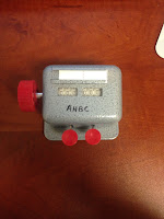
Bellow are some photos of the equipment I used and the cell plate I counted. The first picture is a counter. The second is one of the cell plates counted. It has six wells and each well had to be counted individually.


Also, these are the final counts for each plate. It's a lot of number and columns I know, but don't stress (I mean not that you really were anyways) I'll provide an analysis of the data next time.


Wednesday, March 16, 2016

March 3rd

Thursday, March 3rd marked the second day of my fourth week and the beginning of the second month of the senior research project. I was now fully acclimated to the lab. I truly enjoyed it and felt at home in the lab.

In the cell culture room, I was able to plate cells for the PC3 clonogenic assay. Dr. Lacombe did part of it, but I was able to do a hefty portion. Though I was significantly slower than Dr. Lacombe, I completed it successful. Along the way there were some slip ups. At times I filled the pipette too much or had to throw away a pipette because it touched something that was not sterile. Even though, I completed it and felt accomplished. In addition, I also had the opportunity to freeze cells and changed the medium in flasks.

All in all, as Dr. Lacombe gives me more opportunity to complete tasks in the lab I become more confident and comfortable with lab work.

February 29th

Unfortunately I have run out of swimming and water metaphors, but I have plenty of interesting things to write about my internship. Monday February 29th was the beginning of my fourth week. In the cell culture room, we changed the medium in the 22Rv1 cell line clonogenic assay and trypsinized the DU145 cell line flasks. I also learned a new procedure- how to freeze cells.  By freeze I do not mean place them in the freezer of a refrigerator (though we do do that at one point in the protocol) I mean place them in liquid nitrogen. Freezing cells is an extremely delicate process and requires you to work quickly, so for the first time I watched Dr. Lacombe complete the procedure.

Once we were done in the cell culture room, we washed the cell plates of the DU145 clonogenic assay. This involves dying the cells so you can count the colonies. Once the cells have been dyed and the plates have been washed, they must be left to dry for two days.

Finally Able to Swim Again.

The third week of the internship was the week everything clicked; I was finally able to swim again.

Now that I was using the right hand for pipetting (which happens to be my right hand) the lab work went more smoothly. Though now I had to get acclimated to the shift in placement of everything. When working in the lab, a major key to have a successful experiment (this is something you will not learn from DJ Khaled) is to minimize contamination as much as humanly possible. And I say humanly possible because nobody is perfect. Ways to do this is by not using the same pipette for two different cell lines; not using the pipette if it touches something that is not considered sterile;  not passing the pipette over lids or open bottles; and many more procedures that will only really benefit you to know if you work in a lab so I won't bore you.

In addition to being one step closer to being a pipetting "master", I finally truly understood the goal of the projects. Not that I did not understand before, I did. I just had not my "aha" moment. The first couple weeks were like my first time in the ocean; constantly getting caught in wave after wave and having no idea how to prevent it from happening again. This week, though, the lightbulbs went off. I understood the purpose of each experiment, the significance of the projects, what the expected outcome, and how to complete the lab work.

Also, on the Friday February 25th, I was introduced to a new experiment- the REDI-Dx Assay (CLPA Assay). This is a genetic analysis test. We completed this for the DU145 cell lines and analyzed approximately 20 genes.

Watch Out for That Wave!

The second week of the internship was a blur. Though I was acclimated to the lab and knew what I was going to be working on, there was so much more to do than I expected. If you are a person who loves training, then this is definitely the field for you. I do not mean learning how to use machines and lab equipment; I mean reading paragraph after paragraph about different safety protocols and regulations. Granted, it was interesting at times and definitely gave me a better understanding of the how the lab worked, but man was there a lot of it. Besides training, my second week consisted of learning more procedures; this time I learned how to seed cells for a clonogenic assay. Basically, I took cells from flasks and put them on a six well plate. I'll provide pictures of a flask and clonogenic assay in a later post.

One of the most exciting part of the week was going to the Mayo Clinic in Scottsdale to have the cells irradiated. The irradiator is located in one of the research buildings on the left side of the hospital. Operating the machines is fairly straightforward. Since lead is build into it we did not have to wear personal protective equipment or go to another room. We did wear gloves for precautionary reasons, but the process was simple. Dr. Lacombe placed the cell plate in the respective place and the radiation technician, Kevin, inputed the number of seconds the cells would be needed to irradiated to get the correct dosage (2Gy, 4Gy, 6Gy, 8Gy, or 10Gy) and turned on the X-rays. Though we were there for only 30 minutes, it was exciting to be able to witness the process and helped me to better understand what the project was about.

All in all, my second week was still an informative week for me. I continued to learn the ropes of the lab and learned what to do and what not to do. For example, when pipetting use your dominant hand not your non-dominant hand just because the person who taught you uses that hand. Other than being hit with a tsunami of training, my second week was a fun one. 

Tuesday, March 15, 2016

Diving in Head First

Hello, my name is Nandini Sharma and welcome to my blog for the Senior Research Project. I will be working at the University of Arizona Center for Applied NanoBioscinece and Medicine with Dr. Jerome Lacombe. I will be in the lab on Monday, Thursday, and Friday of every week.

Going into my first week of the Senior Research Project I had only the slightest idea of what I would be doing. The only solid facts I knew, or at least thought, were that I was going to have to change my proposal and I was probably going to get very little hands-on time with equipment and the experiment. Alas, as I found out on my first day, the second assumption was absolutely, inexplicably wrong.

On my first day I was like a kid thrown head first into the pool. From the moment I got to the lab I was put to work. First I learned what project, or projects, I would be working on: This is a project studying the effects of radiation on prostate cancer. There are two studies. in the first study,  we will determine the cellular response of three prostate cancer cell lines (PC3, DU145, and 22Rv1) after single or fractionated doses of radiation. The second study focuses on identifying radioresistant biomarkers of prostate cancer. For the second study we will only be using the PC3 cell line. After Dr. Lacombe and I went to the cell culture room. This is the room where the magic happens; at least I like to think so. In this room there is a Biosaftey Cabinet, incubator, fridge, centrifuge, microscope, and all other supplies needed to complete experiments. I won't bore you with the details of what exactly went on, but basically, I learned to use the pipettes (from the micropipettes- 1mL or less to the large ones- 32 mL), change medium of the flasks (remove the liquid that the cells grow in and add a fresh one), and "trypsinize" cells ( moving cells to a new flask so you can continue to culture them). Then I got to complete my first experiment- a micro-BCA Protein Assay. All in all, my first day was far more exciting than I thought it would be.

The rest of the week was just as hands on, but not always as fun. The second day I counted cells for the clonogenic assay. I'm not going to lie this is tedious and, honestly, not that exciting. Since the lab does not have the equipment to automatically count the colonies we have to hand count the cells. This means you sit for hours looking into a microscope marking each colony with 50 or more cells with a sharpie. But if you've ever seen a sharpie under a microscope it's not that bad. The next day, Friday, I was able to analyze the data for the clonogenic assay and the micro-BCA Assay. Unfortunately, we had to throw out the data because the experiments were no longer viable, but it was good practice and an opportunity for me to get acclimated to the work I would be doing in the next few weeks.

All in all, my first week was a whirlwind. I went in having know idea what to expect and came out completely in awe of the awesomeness of the lab and thrilled that I had the opportunity to work there. These next few months were going to be amazing.